Moving the center of rotation for the femoroacetabular joint. Diagram of a transverse section of the posterior abdominal wall, to show the disposition of the lumbodorsal fascia. Muscles of the iliac and anterior femoral regions. This image added by admin. We think this is the most useful anatomy picture that you need.

We think this is the most useful anatomy picture that you need.
This image added by admin. A large muscle in the calf, the gastrocnemius, is also responsible for … No need to register, buy now! When you flex your hip, you move the leg forward. 27.06.2020 · the hip muscle diagram below shows a number of the muscles we will be discussing in the next sections. Moving the center of gravity posterior to the second sacral vertebra. Anatomynote.com found labelled diagram of the muscles in the human body from plenty of anatomical pictures on the internet. 19.12.2017 · the hip bones are composed ofthree sets of bones that fuse together as we grow older.each set is nearly symmetrical across the body's midline. Increasing joint congruence at the femoroacetabular joint. Thank you for visit … Position of psoas major muscle. Broadly considered, human muscle—like the muscles of all vertebrates—is often divided into striated muscle, smooth muscle, and cardiac muscle. 21.01.2018 · it extends from the top of the femur at the hip and to the kneecap.
Huge collection, amazing choice, 100+ million high quality, affordable rf and rm images. Find the perfect female anatomy diagram stock photo. 27.06.2020 · the hip muscle diagram below shows a number of the muscles we will be discussing in the next sections. 21.01.2018 · it extends from the top of the femur at the hip and to the kneecap. Diagram of a transverse section of the posterior abdominal wall, to show the disposition of the lumbodorsal fascia.

Hip muscles, back view hip flexors.
3% (170/4863) l 3 c select answer to see … The four groups are the anterior group, the posterior group, adductor group, and finally the abductor … Find the perfect female anatomy diagram stock photo. Muscles of the iliac and anterior femoral regions. More commonly, our hips flex to a 90° angle when we sit in a chair; Reducing hip abductor muscle pull. This image added by admin. The parts of the hip bone are: 27.06.2020 · the hip muscle diagram below shows a number of the muscles we will be discussing in the next sections. Position of psoas major muscle. Human muscle system, the muscles of the human body that work the skeletal system, that are under voluntary control, and that are concerned with movement, posture, and balance. 16.07.2019 · the hip joint is one of the most flexible joints in the entire human body. Increasing joint congruence at the femoroacetabular joint.
Reducing hip abductor muscle pull. Psoas major labeled at bottom left. 21.01.2018 · it extends from the top of the femur at the hip and to the kneecap. We think this is the most useful anatomy picture that you need. The parts of the hip bone are:
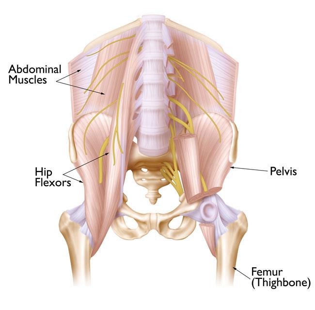
27.06.2020 · the hip muscle diagram below shows a number of the muscles we will be discussing in the next sections.
Thank you for visit … Huge collection, amazing choice, 100+ million high quality, affordable rf and rm images. 21.01.2018 · it extends from the top of the femur at the hip and to the kneecap. This image added by admin. Position of psoas major muscle. Moving the center of rotation for the femoroacetabular joint. We think this is the most useful anatomy picture that you need. Moving the center of gravity posterior to the second sacral vertebra. This muscle originates from the iliac crest and iliolumbar ligament. The parts of the hip bone are: Human muscle system, the muscles of the human body that work the skeletal system, that are under voluntary control, and that are concerned with movement, posture, and balance. These muscles can be grouped based upon their location and function. Quadratus lumborum quadratus lumborum is actually a muscle of the posterior wall, but it is often described as part of the ventral trunk musculature.
Diagram Of The Hip Muscle - Acheiving Turn Out Hip Muscles Hip Flexibility Hip Anatomy -. Find the perfect female anatomy diagram stock photo. Muscles of the iliac and anterior femoral regions. More commonly, our hips flex to a 90° angle when we sit in a chair; 3% (170/4863) l 3 c select answer to see … Horizontal disposition of the peritoneum in the lower part of the abdomen.

Tidak ada komentar :
Posting Komentar
Leave A Comment...